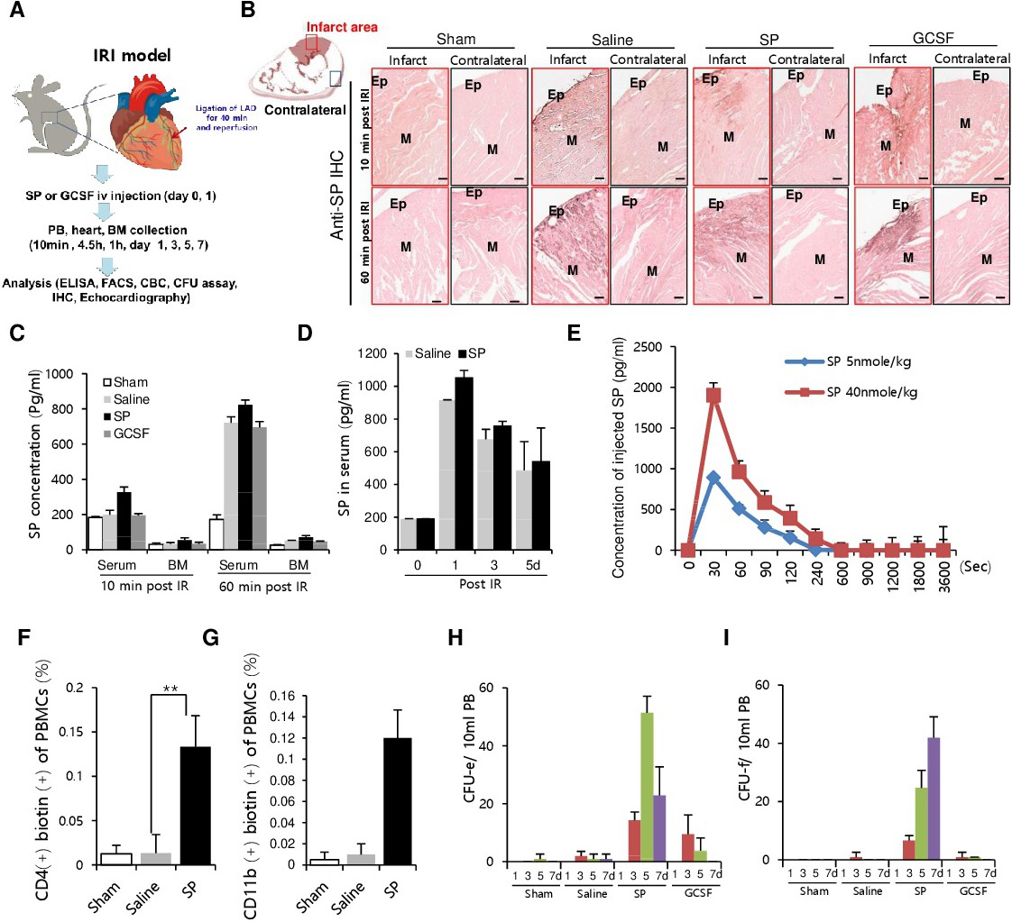
Fig. 2. Detection of endogenous substance P (SP) elevation and mobilization of endothelial progenitor cells (EPCs) and bone marrow mesenchymal stem cells (BMSCs) during ischemia reperfusion injury (IRI). (A) Schematic model of IRI in SD rats and experimental the schedule. SP or granulocyte colony- stimulating factor (GCSF) was intravenously injected immediately after reperfusion. (B) Immunohistochemistry of SP in the paraffin-sectioned cardiac tissue of the infarcted zone and the contralateral side. EP: epicardium, M: myocardium. Scale bar: 100 µm. (C, D) Enzyme-linked immunosorbent assay (ELISA) of SP in the blood and bone marrow (BM) at 10 min and 60 min post- saline, SP, or GCSF injection after IRI, and in the blood on days 1-5 post-IRI. (E) Analysis for pharmacokinetic parameters of SP in the blood. (F, G) At 10 min after Biotin-SP injection in the IRI model, PBMCs were isolated. Biotin-SP bound cells were detected by FITC-streptavidin, CD4+ T cells were determined by APC-conjugated CD4 antibody, CD11b monocytes were determined by APC- conjugated anti-CD11b antibody, and followed by FACS analysis. (H, I) Comparison of CFU-f and CFU- e cell-mobilization kinetics in the sham-operated (n = 6), IRI + saline (n = 6), IRI + SP (n = 6), and IRI + GCSF (n = 6)-injected groups. The data are shown as the mean ± SD. *p<0.05, **p<0.01, ***p<0.001.